Malar Cheek Anatomy
Https Bmendelson Com Au Wp Content Themes Bm Pdf 2008 20surg 20anat 20of 20mid 20cheek Pdf
Index Home

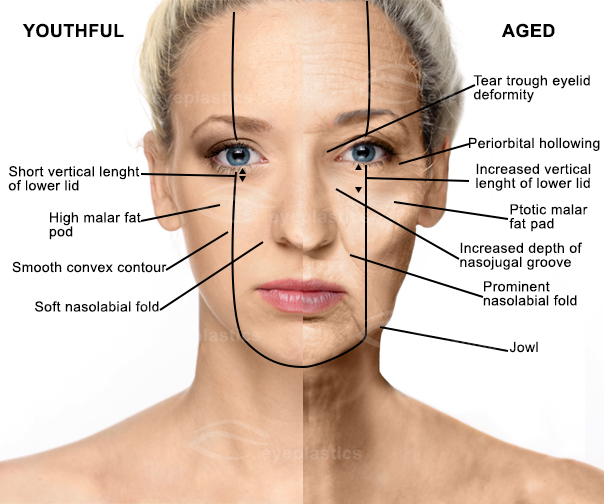
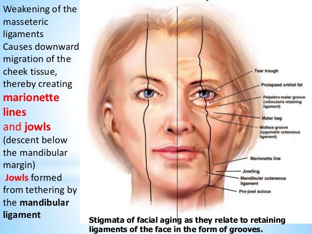
Right Youthful Contours Left Aged Anatomical Findings
Reconstruction Of The Cheek Plastic Surgery Key
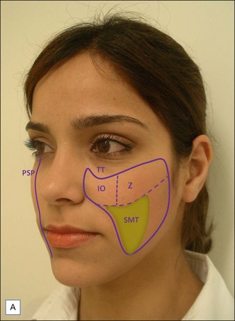
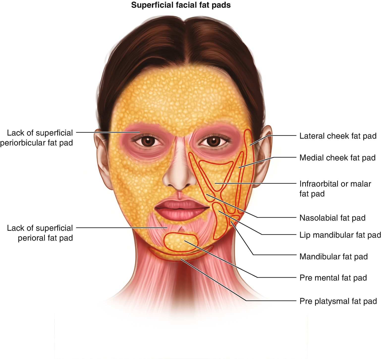
Anatomy And Aging Of Cheek Fat Compartments
Elements Of Morphology Human Malformation Terminology

The anatomy of malar mounds and malar edema is evaluated in a series of 18 fresh cadaver dissections.
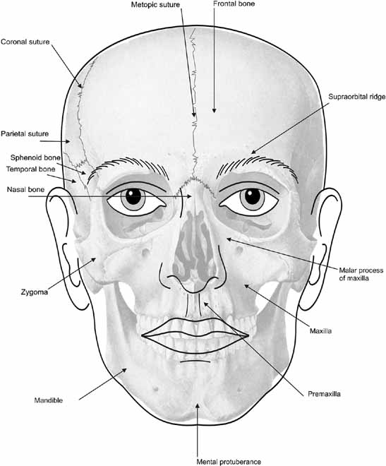
Malar cheek anatomy. Malar the arch of bone beneath the eye that forms the prominence of the cheek cheekbone jugal bone malar bone os zygomaticum zygomatic zygomatic bone jugal point jugale the craniometric point at the union of the frontal and temporal processes of the zygomatic bone. Understanding facial aging skinspirationsskinspirations these two bones join to form the cheekbone. This fascial structure acts as a relatively impermeable barrier that allows tissue edema. Dye injection histologic evaluation and gross anatomic dissection are used to identify a previously unrecognized fascial structure of the lower eyelid and cheek.
The malar septum originates from orbital rim periosteum superiorly and inserts into cheek skin 2 5 to 3 cm inferior to the lateral canthus. It is situated at the upper and lateral part of the face and forms the prominence of the cheek part of the lateral wall and floor of the orbit and parts of the temporal fossa and the infratemporal fossa. The body of the zygomatic bone is roughly quadrangular and has two processes. The cheekbone is a major part of the anatomy of the skull.
One to the frontal and the other to the temporal bone fig. The anatomy of the midcheek has not been satisfacto rily described to adequately explain midcheek aging and malar mounds nor has it suggested a logical approach to their correction or provided sufficient detail for safe sur. The zygomatic bone bridges the facial skeleton to the cranial bones by connecting the. The anatomy of the midcheek has not been satisfactorily described to adequately explain midcheek aging and malar mounds nor has it suggested a logical approach to their correction or provided sufficient detail for safe surgery in this area.

Pdf Local Flaps Cheek And Lip Reconstruction

Pin Em Anatomy And Pose
Three Dimensional Approach Of Cosmetic Patient Aging Gracefully

Aging Face Facial Aging Facial Aesthetics Face Anatomy

Vascular Anatomy Of The Cheek And Lip Download Scientific Diagram
Cheeks Clinical Gate
Fawzy A Fat Compartments And Retaining Ligaments Of The Face
Image Result For Malar Part Of Cheek Estetica Facial E Corporal

Rejuvenation Of The Upper Face I Yimei Search For Aesthetic
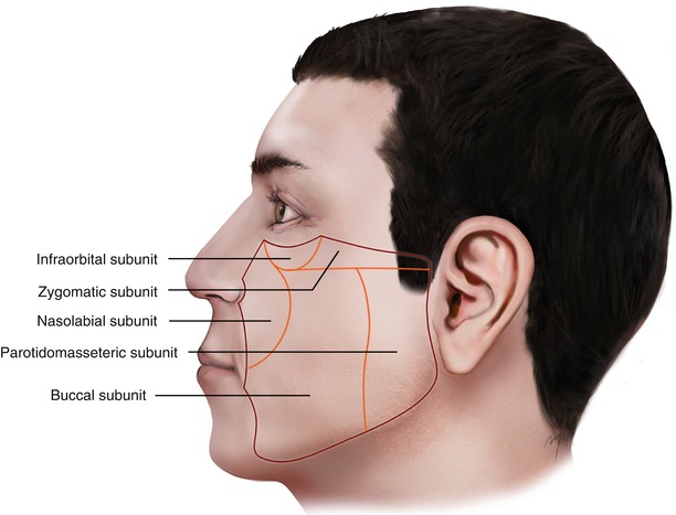
Cheek Plastic Surgery Key
Facial Ultrasound Anatomy For Non Invasive Cosmetic And Plastic

Anatomy Of The Cheek Implications For Soft Tissue Augmentation

Figure 6 16 From I Aesthetic Surgery Of The Face 6 Anatomy Of The
